How EDX Analysis Can Help Identify Contaminants in Your Samples

EDX, or energy-dispersive X-ray spectroscopy, is a chemical microanalysis technique that requires little to no sample preparation and is non-destructive. It is a powerful tool for contamination analysis and industrial forensic science.
Companies like Microvision Labs use EDX to determine elemental composition through a process called electron transitions. An electron residing at a higher energy level sees a vacant space inside an atom and jumps to a lower one, releasing x-rays.
Detecting Microorganisms
Detection of microorganisms is key to food safety, as they can cause contamination and spoilage. Traditional detection methods involve culturing bacteria or other microorganisms using nonselective and selective enrichment techniques, followed by biochemical confirmation.
However, detecting and identifying microorganisms in samples can take time and effort. This is especially true if the sample is complex and contains many components, including microbial and metabolite traces.
Rapid microbial identification is essential to prevent the spread of pathogens and ensure food is safe for consumers. Molecular methods, such as 16S ribosomal RNA sequencing and polymerase chain reaction (PCR), can quickly identify and confirm the presence of bacteria, molds, and yeasts in the sample.
Identifying Contaminants in Metals
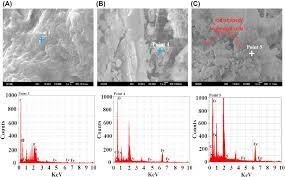
Whether you are trying to identify contaminants in your sample or want to understand how a component will degrade, energy-dispersive X-ray (EDX) analysis can help you. Used with a scanning electron microscope, EDX analysis services like what you can find at microvisionlabs.com provide elemental identification and quantitative composition information on various materials.
EDX is a technique that uses an electron beam of 10-20 keV to bombard a sample with a high-energy X-ray. An energy-dispersive spectrometer then collects those X-rays.
A spectrum is generated with peaks at characteristic energies for each element present. The areas under selected peaks give semi-quantitative elemental composition information.
EDX is a widely-used elemental identification tool for determining the elements present in a sample, their concentration, and their distribution throughout the sample. This information is essential for identifying the chemical structure of a material and is useful for generating composition percentages, which can be correlated to a chemical formula or atomic weight.
Identifying Contaminants in Plastics
Scanning Electron Microscopy (SEM) is a powerful tool for identifying contaminants in various materials. With the addition of an X-ray detector (EDX), SEM becomes an even more powerful tool for identifying contaminants.
EDX is an elemental and composition analysis method that uses characteristic X-rays produced by electron beam irradiation. The energy spectrum of these X-rays gives information about the elements present in the sample, including their concentration and phase.
The X-rays are produced by the electron beam interacting with atoms in the sample. Because of their different, discrete energies, a beam of electrons can knock out an atom’s electrons and produce an X-ray of characteristic wavelength. The SEM then detects the X-rays to reveal chemical information about the sample.
Identifying Contaminants in Food
Identifying contaminants in your samples is vital to help you:
- Assist investigations.
- Avoid further contamination.
- Decide whether a product recall is needed.
- Fulfill legal obligations, reassure consumers, and protect the brand.
EDX analysis can provide a non-destructive, rapid, and accurate method for identifying physical contaminants in food and other materials.
EDX is an electron microscope microanalysis technique that uses high-energy x-rays to produce elemental spectra. These spectral data show the peaks of atoms containing specific elements in the sample.
In the case of EDX, x-rays travel in almost straight lines, which allows the beam to pass through the specimen without interfering with it (this is known as “backscattered electrons”). The backscattered x-rays produce an image on a detector to one side of the sample. The image is then converted into an electron microscope picture. This enables the analysis of samples whose morphology or composition is unsuitable for other analytical techniques.


